Tsinghua University announced on June 12th that its research team captured high-definition continuous footage of human preimplantation embryonic development over five days for the first time globally using advanced long-term live-cell microscopic imaging technology. The team identified the major trigger behind the high incidence of human embryonic developmental arrest. This breakthrough study was published online in the international academic journal Cell.
In vitro fertilization (IVF) has brought new hopes to patients with infertility. Still, the success rate of IVF treatment remains constrained by critical hurdles. Clinical data show that more than half of human fertilized eggs suffer developmental arrest within the five-day window from fertilization to blastocyst formation. These arrested embryos cannot be transferred back into the maternal uterus for implantation, which stands as a primary barrier to successful pregnancy.
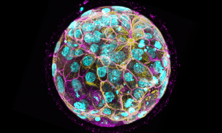
To address this longstanding clinical challenge, a research group led by Dr. Jun Su, Assistant Professor at Tsinghua’s Institute of Biomedical Interdisciplinary Research and Investigator at the National Institute of Biological Sciences, Beijing, independently developed a high-throughput dual-view light-sheet fluorescence microscope. The device enables sustained high-resolution live imaging of embryos and delivers far richer imaging datasets.
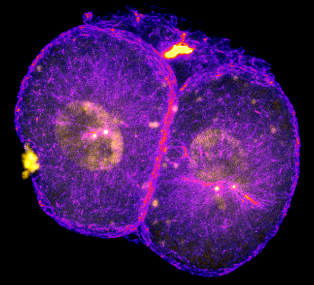
The team systematically analyzed over 2,000 cell division events across more than 150 human and cynomolgus monkey embryos. Their analysis revealed that over 70% of embryos arrested in the first three days of development exhibited spindle abnormalities during the second cleavage stage.
A spindle refers to a spindle-shaped cytoskeletal structure formed during cell division. Dr. Su explained that human embryos undergo repeated cleavage divisions. In each division cycle, spindles are required to evenly segregate chromosomes into two daughter cells. Experimental evidence confirms that spindle defects arising at the second cleavage directly cause chromosomal abnormalities, which trigger embryonic cell cycle arrest within the subsequent three rounds of cell division.
Follow-up investigations further pinpointed centrosomes – intracellular cytoskeleton-organizing centers – as a critical regulator of spindle assembly in human preimplantation embryos. Abnormal centrosome counts directly drive the formation of defective spindles.
Building on these discoveries, researchers treated embryos at the second cleavage stage with a centrosome replication regulatory protein inhibitor at a semi-inhibitory concentration. The intervention raised the proportion of cells with normal centrosome numbers from 40% to 80%, drastically lowering the probability of centrosome replication errors in embryos. Notably, the treatment exerted no adverse effects on embryonic cells with intact centrosome counts.
“This finding holds promising potential for clinical intervention and prevention of early cleavage-stage embryonic arrest”, Dr. Su commented. His team will continue developing innovative microscopic imaging tools to build a more efficient and safe embryonic developmental evaluation system, with the ultimate goal of boosting embryo implantation rates.